
Hormone blood test (men)
Testosterone and related hormones affecting energy, mood, and drive.
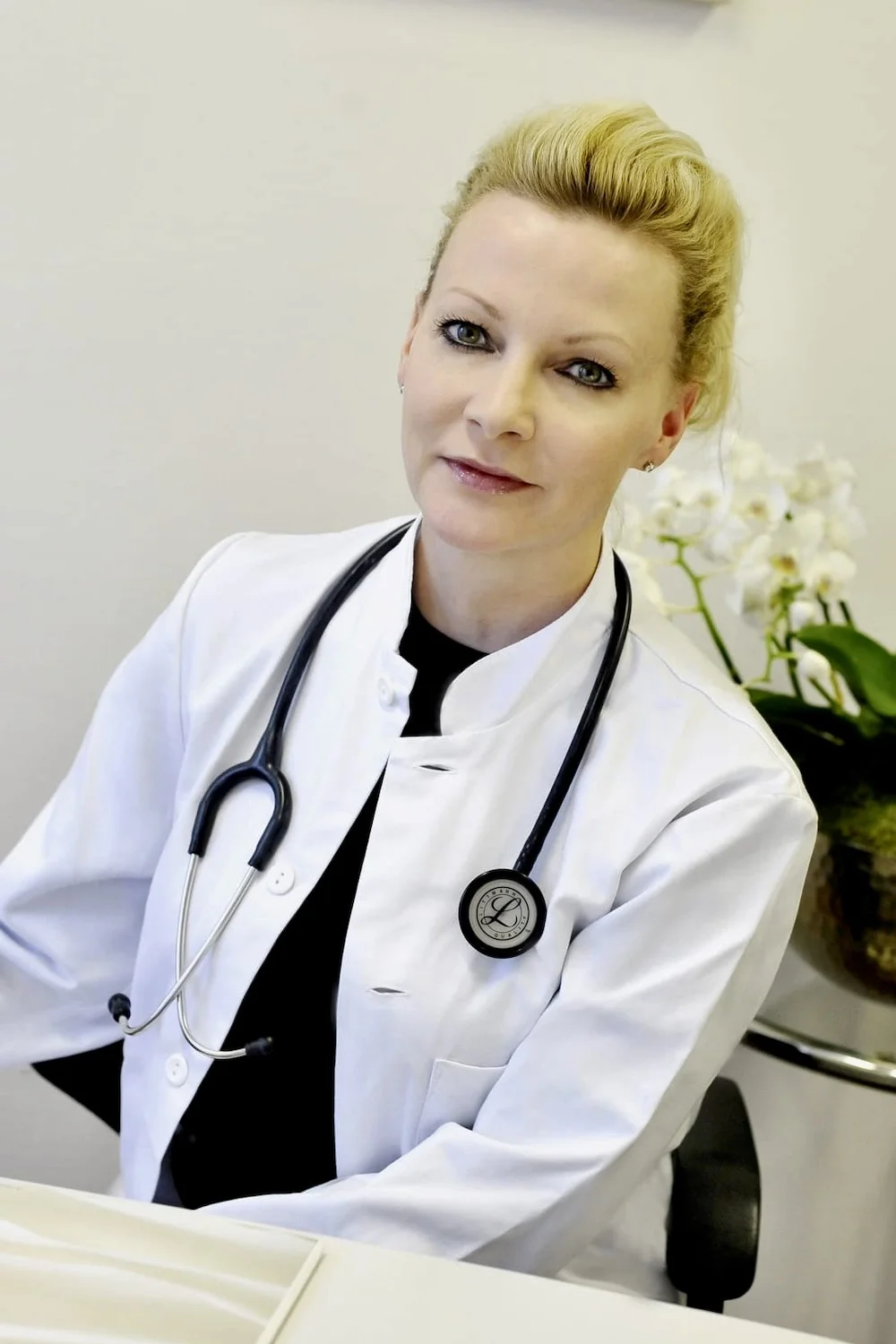
Reviewed by Swiss physicians
Personal action plan
Interactive report
Questions we could help with


TIME NEEDED
15min
Quick appointment at our clinic
HEALTH AREAS
Energy, mood, and vitality
BIOMARKERS TESTED
5
Key hormone markers
Measures testosterone and related hormones affecting energy, mood, and drive.
PREPARATION REQUIRED
Hormones don't need fasting. As an add-on, follow your main blood test's fasting rules. Best in the morning.
FOR WHOM
18-80
Men monitoring hormonal balance
Our blood test packages
Each blood test includes a clear report reviewed by a doctor, starting at CHF 299.

Advanced blood test
Duration: 15 min
Advanced blood test that goes beyond GP coverage. Includes a comprehensive 80+ biomarker blood test.
Reviewed by Swiss physicians
Personal action plan
Add tests or MRI imaging anytime

Your insurer may cover your check-up
Multiple supplementary insurance plans contribute to the cost of our preventive check-ups. We've confirmed coverage with leading Swiss providers. Reimbursement varies by plan.
Three simple steps to your blood test results

Before your appointment
Our team will review additional details so you're fully prepared — no referral needed.

During your appointment
The clinic team welcomes you and takes a small blood sample. Expect your visit to take about 20 minutes from start to finish.

After your appointment
Your blood is analyzed, and within a few days you receive a digital report and personalized health plan from our physicians.
Health reports without the headache
Medical data can be complex. Our interactive reports explain each result in simple terms with clear visuals.


Your recent health check-up provides a detailed overview of your health, showing some very positive developments alongside a few areas that warrant attention. The most notable findings are related to age-appropriate wear-and-tear in your spine.
Made for each other
The hormone blood test can be combined with other Ahead tests during the same visit to maximize health insights.

Full-body MRI
A full-body scan offering detailed insights into major organs for preventive health.

Advanced blood tests
Test 80+ biomarkers, including cholesterol and glucose, for a full health overview.

Omega-3 & 6 blood test
Measure 16 biomarkers to know if your Omega intake meets your body's needs.

Hormone test for women
Test 9 key hormones to clarify symptoms and support cycle health.

Essential-B-vitamins blood test
Check B12 and folate (B9) levels to reveal if deficiencies are behind fatigue or low focus.
~ Shown values are illustrative samples.
Real results from real people
Every health journey is unique. Read how others took control of their health with Ahead.

The experts behind your health journey
Every scan is personally reviewed by one of our renowned radiologists. Their combined expertise ensures you get the most accurate and actionable results.
30,000+
MRI's reviewed over 10+ years

Prof. Dr. med. Edouard Battegay
Emeritus Professor of Internal Medicine at the University of Zurich. Head of the International Center for Multimorbidity and Complexity in Medicine.

Dr. Jacob J. Visser
Radiologist / CMIO. Assistant Professor of value-based imaging at Erasmus University Medical Center.

Prof. Dr. Benedikt Wiestler
Professor of AI for image-guided diagnosis and therapy at the Technical University of Munich.

Prof. Dr. med. Michael Fischer
Professor of Radiology at the University of Zurich, abdominal and musculoskeletal specialist.

Prof. Dr. med. Olivio Donati
Professor of Radiology at the University of Zurich, oncology specialist and leading Swiss prostate expert.
Dr. med Anna Erat, MD, PhD
Harvard Medical School-trained prevention expert. Faculty position at the University of St. Gallen Executive School.
